
Caitlyn Coleman
Introduction
Archaeological research has had a considerable impact on modern discoveries. Studies on skeletal remains and mummified samples led to improvements in the research process for modern infections. Ancient DNA research has its challenges with controlling contamination and the fragmentation of the samples, but improvements in methodology such as Uracil DNA glycosylase (UDG) treatments [Llamas et al, 2016] have mitigated damage in some regard. However, it is necessary to consider more than just DNA, but a holistic approach that includes proteomics, metabolomics, and genomics. The objective of this literature review is to synthesize the advances in these three studies with an emphasis on infectious diseases. There is specific attention paid to the bacterium Yersinia pestis, and the Black Death.
Proteomics in Archaeology
Applications within Mummification
Protein networks within ancient DNA contain vital information to better understand infectious diseases. The identification of various proteins and the discovery of new putative proteins furthers archeological research. The presence of certain proteins can indicate an immune response or point to inflammation, which demonstrates possible infection [Jones et al, 2016]. The most abundant category in a study using ancient Egyptian mummies was anatomical structure development, specifically collagens and keratins [Jones et al, 2016]. Another study on a Korean mummy used genomic analysis to pinpoint four open reading frames (ORFs) that aligned with the hepatitis B virus [Bar-Gal et al, 2012] . The strain of hepatitis B virus that was mummy-derived was compared to modern DNA to establish how long the virus had been in East Asia and how it had evolved over thousands of years [Bar-Gal et al, 2012]. Through bioinformatics analysis, specific primer sequences found in Inca mummy samples were matched to genomes in the phylum Actinobacteria [Corthals et al, 2012]. Gel electrophoresis confirmed the results with two lanes producing positive results that matched Mycobacterium tuberculosis [Corthals et, 2012]. These studies on mummies from different countries has increased the knowledge of pathogen evolution and the spread of infectious diseases in both past and present times.
Fifteen immune response proteins, three stress-response proteins, and twenty-six metabolic proteins were found through proteomics analysis of muscle tissue biopsies of an adult mummy [Jones et al, 2016]. These metabolic proteins included enzymes such as Glyceraldehyde 3-phosphate dehydrogenase (GAPDH) and Amylase which both connect to inflammation [Jones et al, 2016]. Remains alone have not been able to reveal the presence of disease, but in combination with aDNA analysis, stronger results have been produced.
Microbiological Application within the dental calculus
The dental calculus provides insights to the microbiome of ancient civilizations. It allows researchers to document disease throughout history. Specifically, dental plaque holds hundreds of types of bacteria [Preus et al, 2011]. While it is noted that there is stability in the oral microbiome, it is one of the most accessible areas for pathogens to enter the body [Preus et al, 2011]. Germs can easily be transferred from hard surfaces to people’s hands, but from here, they must find a way inside. One way is through improper handwashing, or its absence altogether, and then touching one’s face or food. Deadly infections can be transmitted this way, and even in death, the killer microorganism can be found in the victim’s saliva [Preus et al, 2011]. It was found that the protein taxonomy has variability, and certain categories are expected to show up more often [Mackie et al, 2017]. While pathogenic bacteria were identified, that was only one of three sources of bacterial proteins. [Mackie et al, 2017]. Other sources included opportunistic and commensal species of bacteria. [Mackie et al, 2017]. New studies have the potential to disclose mechanisms involved in the evolution of microbiome bacteria [Weyrich et al, 2015]. The dental calculus preserves the pathogen of that time.
Dietary Application within the dental calculus
Dental calculus also provides information on the diet of the specimen. It can narrow down specific food groups that were consumed more often than others. Fragments of the food consumed are preserved in the dental calculus deposit along with biomolecules and diagnostic proteins [Hendy et al, 2018]. Many diseases are transmitted from animals to humans, so it is important to note what species were a main food source during the time period of study. The microfossils within dental calculus hold details about the evolution of various plants and animals that made up the dietary behavior of various cultures [Weyrich et al, 20 15]. The dental calculus is a record of the dietary habits of the people it is sampled from.
Application using Yersinia pestis and Yersinia pseudotuberculosis
In Yersinia pestis, tracking the activity of the urease enzyme led to a discovery that explained how the plague evolved over time. This enzyme played a key role in the transmission of the bacteria throughout Europe by fleas [Chouikha and Hinnebusch, 2014]. Urease is toxic to fleas, and its deletion increased the potential for transmission, hence the mutation was favored in Yersinia pestis [Chouikha and Hinnebusch, 2014]. In a past study the results proved that the presence of functional urease is a requirement if the bacteria Yersinia pseudotuberculosis was to have any toxicity in fleas [Chouikha and Hinnebusch, 2014]. Urease enzymes have further explained the evolution of the Black Death.
Metabolomics in Archaeology
Infectious Disease Application
Metabolic processes play an important role in the study of infectious diseases. They are interrupted by the infection, and that disruption impacts the end products or metabolites [Ghost et al, 2018]. The sensitivity of these pathways to changes in the environment, or increased stress make the infection progress easily visible [Ghost et al, 2018]. In the case of malaria, the effect on the host’s ability to utilize glucose starts a chain reaction that lowers the production of creatine, increases the levels of creatine phosphokinase (CPK), and trickles down to a decrease in phosphocreatine synthesis [Ghost et al, 2018]. In addition, metabolites from Brown propolis used in a biofilm were successful in decreasing the amount of Staphylococcus aureus and T. vaginalis [Dembogurski et al, 2018]. Phenylpropanoic acids and flavonoids are two significant compounds that caused the decrease in bacterial activity [Dembogurski et al, 2018]. Metabolites are a way to follow infectious disease through the biological systems of their host.
Lipidomics Subset
Lipidomics concentrates on how lipids interact with the metabolites of metabolomics.
Two lipids that hold great significance in the study of infectious diseases are phosphatidylcholine (PC) and phosphatidylethanolamine (PE) [Wang et al, 2019]. Phospholipids are also important to monitor as any fluctuation in their levels detect changes in an organisms’ overall functions [Qin et al, 2016]. Analysis of lipids often involves either radioactive or fluorescent labeling to keep track of the sample [Lam and Shui, 2013]. It also requires mass spectrometry and chromatography to have a full lipidomic approach [Lam and Shui, 2013]. Lipid interaction is another method of tracing the spread of an infection through the host organism.
Genomics in Archaeology
General Genomics
Genomics is the study of an organism’s genes in their entirety: the genome. There are complications that arise when dealing with ancient DNA genomes that make it difficult to show the diversity in various pathogens responsible for past outbreaks [Andam et al, 2016]. Uracil selection (otherwise known as U selection) is a new method that has shown promise in decreasing contamination in ancient genome research [Gansauge and Meyer, 2014].
However modern outbreaks such as the Ebola and Cholera outbreaks have benefitted from genome sequencing [Andam et al, 2016]. Genes hold much more than genetic material, they have the potential to improve infectious disease research of both ancient and modern pathogens.
Applications using Yersinia pestis
Genomic analysis of Yersinia pestis allows researchers to better understand how it spread so rapidly across Europe. Three historical genomes studied contained specific single-nucleotide polymorphisms (SNPs), within the pabA and NCa genes. [Spyrou et al, 2016]. These genomes ended up on the same phylogenetic branch, even though they originated from Barcelona, Bolgar City, and Ellwangen [Spyrou et al, 2016]. It was found that when comparing Yersinia pestis to Yersinia pseudotuberculosis, isolates of pestoides and microtus were more closely related to Yersinia pestis. [Atchman et al, 2004]. The genome of Yersinia pestis provided more insight to how it caused multiple pandemics.
Current Methodologies Flowchart
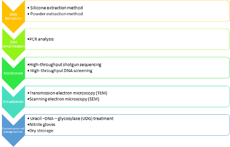
Significant Research Questions
After extensive review it was clear there is commonality among the research questions being pursued. These common questions include:
- How can contamination be minimized in archaeological research?
- What is the most effective combination of treatments to most accurately study ancient DNA?
- What can archaeological research reveal to improve modern studies?
CONCLUSION
In this review, proteomics, genomics, and metabolomics, were outlined as they relate to infectious diseases, and specifically Yersinia pestis. The current methodologies and significant research questions were compiled. None of these disciplines are sufficient alone, but a combination of the three yields more comprehensive research. Proteomics and metabolomics allow a closer look at the impact of infectious diseases on biological systems. Genomics and genome sequences track the spread of these diseases and compare different strains.
BIBLIOGRAPHY
Achtman, M., Morelli, G., Zhu, P., Wirth, T., Diehl, I., Kusecek, B., … Keim, P. (2004). Microevolution and history of the plague bacillus, Yersinia pestis. Proceedings of the National Academy of Sciences, 101(51), 17837–17842. doi: 10.1073/pnas.0408026101
Achtman, M. (2016). How old are bacterial pathogens? Proceedings: Biological Sciences, 283(1836), 1-10. Retrieved March 19, 2020, from http://www.jstor.org/stable/24763436
Andam, C. P., Worby, C. J., Chang, Q., & Campana, M. G. (2016). Microbial Genomics of Ancient Plagues and Outbreaks. Trends in Microbiology, 24(12), 978–990. doi: 10.1016/j.tim.2016.08.004
Bar-Gal, G. K., Kim, M. J., Klein, A., Shin, D. H., Oh, C. S., Kim, J. W., … Shouval, D. (2012). Tracing hepatitis B virus to the 16th century in a Korean mummy. Hepatology, 56(5), 1671–1680. doi: 10.1002/hep.25852
Bos, K. I., Herbig, A., Sahl, J., Waglechner, N., Fourment, M., Forrest, S. A., … Poinar, H. N. (2016). Author response: Eighteenth century Yersinia pestis genomes reveal the long-term persistence of an historical plague focus. doi: 10.7554/elife.12994.011
Bos, K. I., Schuenemann, V. J., Golding, G. B., Burbano, H. A., Waglechner, N., Coombes, B. K., … Krause, J. (2011). A draft genome of Yersinia pestis from victims of the Black Death. Nature, 478(7370), 506–510. doi: 10.1038/nature1054
Chouikha, I., & Hinnebusch, B. (2014). Silencing urease: A key evolutionary step that facilitated the adaptation of Yersinia pestis to the flea-borne transmission route. Proceedings of the National Academy of Sciences of the United States of America, 111(52), 18709-18714. Retrieved March 16, 2020, from http://www.jstor.org/stable/43278909
Corthals, A., Koller, A., Martin, D. W., Rieger, R., Chen, E. I., Bernaski, M., … Dávalos, L. M. (2012). Detecting the Immune System Response of a 500 Year-Old Inca Mummy. PLoS ONE, 7(7). doi: 10.1371/journal.pone.0041244
Dembogurski, D. S. D. O., Trentin, D. S., Boaretto, A. G., Rigo, G. V., Silva, R. C. D., Tasca, T., … Silva, D. B. (2018). Brown propolis-metabolomic innovative approach to determine compounds capable of killing Staphylococcus aureus biofilm and Trichomonas vaginalis. Food Research International, 111, 661–673. doi: 10.1016/j.foodres.2018.05.033
Drancourt, M., & Raoult, D. (2004). Molecular detection of Yersinia pestis in dental pulp. Microbiology, 150(2), 263–264. doi: 10.1099/mic.0.26885-0
Feldman, M., Harbeck, M., Keller, M., Spyrou, M. A., Rott, A., Trautmann, B., … Krause, J. (2016). A High-CoverageYersinia pestisGenome from a Sixth-Century Justinianic Plague Victim. Molecular Biology and Evolution, 33(11), 2911–2923. doi: 10.1093/molbev/msw170
Gansauge, M.-T., & Meyer, M. (2014). Selective enrichment of damaged DNA molecules for ancient genome sequencing. Genome Research, 24(9), 1543–1549. doi: 10.1101/gr.174201.114
Ghosh, S., Pathak, S., Sonawat, H. M., Sharma, S., & Sengupta, A. (2018). Metabolomic changes in vertebrate host during malaria disease progression. Cytokine, 112, 32–43. doi: 10.1016/j.cyto.2018.07.022
Gilbert, M. T. P., Cuccui, J., White, W., Lynnerup, N., Titball, R. W., Cooper, A., & Prentice, M. B. (2004). Absence of Yersinia pestis-specific DNA in human teeth from five European excavations of putative plague victims. Microbiology, 150(2), 341–354. doi: 10.1099/mic.0.26594-0
Hendy, J., Warinner, C., Bouwman, A., Collins, M. J., Fiddyment, S., Fischer, R., Hagan, R., Hofman, C. A., Holst, M., Chaves, E., Klaus, L., Larson, G., Mackie, M., Mcgrath, K., Mundorff, A. Z., Radini, A., Rao, H., Trachsel, C., Velsko, I. M., & Speller, C. F. (2018). Proteomic evidence of dietary sources in ancient dental calculus. Proceedings of the Royal Society B: Biological Sciences, 285(1883), 1–10. https://doi-org.ezproxy.lib.usf.edu/10.1098/rspb.2018.0977
Jones, J., Mirzaei, M., Ravishankar, P., Xavier, D., Lim, D., Shin, D., . . . Haynes, P. (2016). Identification of proteins from 4200-year-old skin and muscle tissue biopsies from ancient Egyptian mummies of the first intermediate period shows evidence of acute inflammation and severe immune response. Philosophical Transactions: Mathematical, Physical and Engineering Sciences,374(2079), 1-19. Retrieved March 16, 2020, from http://www.jstor.org/stable/26114763
Karamanou, M., Panayiotakopoulos, G., Tsoucalas, G., Kousoulis, A. A., & Androutsos, G. (2012). From miasmas to germs: a historical approach to theories of infectious disease transmission. Le Infezioni in Medicina, 20(1), 58.
Lam, S. M., & Shui, G. (2013). Lipidomics as a Principal Tool for Advancing Biomedical Research. Journal of Genetics and Genomics, 40(8), 375–390. doi: 10.1016/j.jgg.2013.06.007
Llamas, B., Valverde, G., Fehren-Schmitz, L., Weyrich, L. S., Cooper, A., & Haak, W. (2016). From the field to the laboratory: Controlling DNA contamination in human ancient DNA research in the high-throughput sequencing era. STAR: Science & Technology of Archaeological Research, 3(1), 1–14. doi: 10.1080/20548923.2016.1258824
Mackie, M., Hendy, J., Lowe, A. D., Sperduti, A., Holst, M., Collins, M. J., & Speller, C. F. (2017). Preservation of the metaproteome: variability of protein preservation in ancient dental calculus. Science & Technology of Archaeological Research, 3(1), 58–70. https://doi.org/10.1080/20548923.2017.1361629
Marciniak, S. (2016). Chapter 6 – Hunting for Pathogens: Ancient DNA and the Historical Record. In Beyond the Bones(pp. 81–100). Academic Press. doi: https://doi.org/10.1016/B978-0-12-804601-2.00006-5
Preus, H. R., Marvik, O. J., Selvig, K. A., & Bennike, P. (2011). Ancient bacterial DNA (aDNA) in dental calculus from archaeological human remains. Journal of Archaeological Science, 38(8), 1827–1831. doi: 10.1016/j.jas.2011.03.020
Qin, L., Zhang, Z., Guo, M., Zhang, Q., Wang, Q., Lu, Z., … Gao, X. (2016). Plasma metabolomics combined with lipidomics profiling reveals the potential antipyretic mechanisms of Qingkailing injection in a rat model. Chemico-Biological Interactions, 254, 24–33. doi: 10.1016/j.cbi.2016.05.022
Rasmussen, S., Allentoft, M. E., Nielsen, K., Orlando, L., Sikora, M., Sjögren, K.-G., … Willerslev, E. (2015). Early Divergent Strains of Yersinia pestis in Eurasia 5,000 Years Ago. Cell, 163(3), 571–582. doi: 10.1016/j.cell.2015.10.009
Reboul, A., Lemaître, N., Titecat, M., Merchez, M., Deloison, G., Ricard, I., . . . Sebbane, F. (2014). Yersinia pestis Requires the 2-Component Regulatory System OmpR-EnvZ to Resist Innate Immunity During the Early and Late Stages of Plague. The Journal of Infectious Diseases, 210(9), 1367-1375. Retrieved March 16, 2020, from http://www.jstor.org/stable/43708068
Rohland, N., Harney, E., Mallick, S., Nordenfelt, S., & Reich, D. (2015). Partial uracil–DNA–glycosylase treatment for screening of ancient DNA. Philosophical Transactions of the Royal Society B: Biological Sciences, 370(1660), 20130624. doi:10.1098/rstb.2013.0624
Rohland, N., Harney, E., Mallick, S., Nordenfelt, S., & Reich, D. (2015). Partial uracil–DNA–glycosylase treatment for screening of ancient DNA. Philosophical Transactions of the Royal Society B: Biological Sciences, 370(1660), 20130624. doi: 10.1098/rstb.2013.0624
Schuenemann, V. J., Bos, K., Dewitte, S., Schmedes, S., Jamieson, J., Mittnik, A., … Poinar, H. N. (2011). Targeted enrichment of ancient pathogens yielding the pPCP1 plasmid of Yersinia pestis from victims of the Black Death. Proceedings of the National Academy of Sciences, 108(38). doi: 10.1073/pnas.1105107108
Shapiro, B., Barlow, A., Heintzman, P. D., Hofreiter, M., Paijmans, J. L. A., & Soares André E. R. (2019). Ancient Dna: methods and protocols. New York, NY: Humana Press.
Skoglund, P., Storå, J., Götherström, A., & Jakobsson, M. (2013). Accurate sex identification of ancient human remains using DNA shotgun sequencing. Journal of Archaeological Science, 40(12), 4477–4482. doi: 10.1016/j.jas.2013.07.004
Spyrou, M. A., Tukhbatova, R. I., Feldman, M., Drath, J., Kacki, S., Beltrán De Heredia, J., … Krause, J. (2016). Historical Y. pestis Genomes Reveal the European Black Death as the Source of Ancient and Modern Plague Pandemics. Cell Host & Microbe, 19(6), 874–881. doi: 10.1016/j.chom.2016.05.012
Wagner, David & Klunk, Jennifer & Harbeck, Michaela & Devault, Alison & Waglechner, Nicholas & Sahl, Jason & Enk, Jacob & Birdsell, Dawn & Kuch, Melanie & Lumibao, Candice & Poinar, Debi & Pearson, Talima & Fourment, Mathieu & Golding, Brian & Riehm, Julia & Earn, David & Dewitte, Sharon & Rouillard, Jean-Marie & Grupe, Gisela & Poinar, Hendrik. (2014). Yersinia pestis and the Plague of Justinian 541-543 AD: A genomic analysis. The Lancet infectious diseases. 14. 10.1016/S1473-3099(13)70323-2.
Wang, R., Li, B., Lam, S. M., & Shui, G. (2019). Integration of lipidomics and metabolomics for in-depth understanding of cellular mechanism and disease progression. Journal of Genetics and Genomics. doi: 10.1016/j.jgg.2019.11.009
Weyrich, L. S., Dobney, K., & Cooper, A. (2015). Ancient DNA analysis of dental calculus. Journal of Human Evolution, 79, 119–124. doi: 10.1016/j.jhevol.2014.06.018
Categories: Archaeology, featured, Genomics, Literature Review, Metabolomics, Proteomics
